To Shield or Not to Shield: Rethinking Patient Protection in Dentomaxillofacial Radiography
Lead aprons and thyroid collars were introduced when dental X-ray systems delivered higher doses, and beam control was limited. That rationale has narrowed. This article examines why routine shielding is being reconsidered in dentomaxillofacial radiography, where evidence still diverges, and what actually reduces dose in modern practice.
Lead aprons and thyroid collars became standard protective tools at a time when dental X-ray systems produced higher radiation exposures and beam control was limited. Imaging technology has changed substantially since then, and so has the evidence supporting how patient protection should be applied.
Today, the clinical question is no longer whether shielding should be used automatically. It is whether shielding improves or compromises the examination in a given clinical setting.
Shielding decisions now intersect with three inseparable domains:
clinical guidance
imaging physics
regulatory policy
A modern shielding policy must reflect differences among imaging modalities and patient populations rather than defaulting to a single rule for every study.
Key Clinical Takeaways
Modern shielding decisions in dental radiography are no longer based on routine use of lead aprons or thyroid collars. They are based on modality, positioning reliability, and patient-specific considerations.
Routine patient shielding is no longer recommended for intraoral, panoramic, cephalometric, or CBCT imaging when modern equipment and positioning protocols are used.
Field-of-view selection is the single most important dose-control factor, particularly in CBCT imaging.
Thyroid collars may be reasonable in selected intraoral examinations, particularly when placement does not interfere with positioning or image quality.
Routine thyroid collar use is generally discouraged in panoramic and cephalometric imaging, where shielding frequently enters the field of view.
In CBCT imaging, shielding is not a primary dose-control method. Justification, field-of-view selection, and exposure optimization determine most of the radiation dose.
Pediatric imaging requires careful judgment rather than automatic shielding.
During pregnancy, fetal exposure from dental imaging is extremely low and is driven primarily by internal scatter rather than direct beam exposure.
Preventing repeat exposures reduces cumulative dose more effectively than shielding alone.
Why Shielding Recommendations in Dentistry Are Changing
Recommendations regarding patient shielding are evolving because the conditions that originally justified routine shielding have changed. Modern dental imaging operates under different exposure levels, equipment capabilities, and quality standards than those that existed when shielding first became routine.
Historically, radiation dose reduction relied heavily on physical barriers because early imaging systems required longer exposure times and delivered broader radiation beams. Film-based imaging demanded higher exposure levels, and beam restriction tools were limited. In that environment, shielding provided meaningful protection.
Today, most dose reduction occurs through improvements in imaging systems rather than through shielding alone.
Historical Role of Patient Shielding in Dental Radiography
Patient shielding became standard in the mid-20th century, when diagnostic imaging systems delivered substantially higher radiation exposures than those used today. Early dental radiographic systems used broad beams, longer exposure times, and film receptors that required higher radiation levels to produce diagnostic images.
Lead aprons and thyroid collars were therefore introduced as practical protective tools. They provided a physical barrier between radiosensitive tissues and the projected beam path at a time when other dose-control methods were limited.
By the 1970s, shielding had become standard practice across both medical and dental radiography. Regulatory requirements reinforced its use, and training programs emphasized visible protection as a central safety measure.
That historical context remains important. Many current expectations about shielding are rooted in conditions that no longer reflect modern imaging technology.
How Advances in Imaging Technology Changed Radiation Exposure
The most significant reductions in radiation exposure in dental imaging have resulted from improvements in equipment and technique rather than from shielding.
Key advances include:
transition from film to digital receptors
use of faster film speeds
improved filtration
shorter exposure times
rectangular collimation
optimized exposure settings
Digital imaging systems require substantially less radiation to produce diagnostic-quality images than earlier film-based systems. Rectangular collimation alone has been shown to reduce patient exposure by more than 40% compared with round collimation.
As radiation levels decreased, the relative contribution of shielding also changed. When tissues outside the projected beam path already receive minimal exposure, the additional benefit of shielding becomes proportionally smaller.
Modern radiation protection in dentistry now depends primarily on:
justified imaging
beam restriction
positioning accuracy
optimized exposure parameters
Shielding remains part of the discussion — but it is no longer the primary method of dose reduction.
Why the Topic Remains Controversial
The shielding debate persists not because the science is unclear, but because different questions are being evaluated at the same time.
Some studies examine whether shielding reduces measured radiation dose under controlled conditions. Others evaluate whether shielding improves the overall clinical examination once positioning accuracy, image quality, and repeat exposure risk are considered. These are not equivalent outcomes. A strategy that reduces dose in a laboratory setting may still perform poorly in clinical practice if it interferes with positioning or increases retake rates.
This distinction explains why professional recommendations do not always appear uniform, even when based on similar scientific data.
Similar conclusions have been reached in broader radiologic practice. The American Association of Physicists in Medicine (AAPM) recommended discontinuing routine gonadal and fetal shielding in medical imaging, noting that shielding may interfere with image acquisition and does not protect against internally scattered radiation.
International recommendations remain less uniform. Several published reviews report measurable reductions in thyroid dose when shielding is used under controlled conditions, particularly in CBCT imaging. However, those same studies consistently show that factors such as field-of-view selection produce substantially larger differences in dose than shielding alone.
This divergence reflects differences in emphasis rather than disagreement about basic radiation physics. Some guidance prioritizes measurable dose reduction, while others prioritize reliability of image acquisition and avoidance of repeat exposure.
Evidence Supporting Both Perspectives
Dosimetry studies demonstrate that shielding can reduce absorbed dose when conditions are carefully controlled. In CBCT phantom investigations, shielding has been shown to reduce absorbed dose by approximately 26% to 53%, depending on scan parameters and positioning. At the same time, variation in field-of-view selection has been shown to produce dose differences ranging from 8% to more than 600%, confirming that scan design remains the dominant factor in dose management.
These findings reinforce an important distinction: shielding may reduce dose, but its relative impact is often smaller than that achieved through appropriate field-of-view selection and exposure optimization.
Clinical workflow considerations introduce additional complexity. When shielding enters the field of view, it may obscure anatomy or produce artifacts that compromise interpretation. In such cases, repeat imaging may be required, resulting in greater cumulative exposure than the shielding would have prevented.
For this reason, shielding decisions must be evaluated not only in terms of measured dose reduction, but also in terms of image reliability and retake risk.
Why the Debate Persists in Clinical Practice
The persistence of the shielding debate is not purely scientific — it is cultural and practical.
For decades, visible shielding became synonymous with safety in dental imaging. Patients expect it. Staff are accustomed to it. In many regions, regulations were written when earlier technology justified routine shielding, and some of those rules remain in place even as professional guidance evolves.
There is also an understandable ethical dimension. Radiation risk, even at low levels, carries emotional weight. Clinicians may prefer to retain shielding practices because they appear protective, even when evidence suggests the benefit is minimal in modern systems.
Finally, uncertainty is amplified by the fact that shielding is not universally ineffective. Under certain conditions — particularly when radiosensitive tissues lie close to the projected beam path — shielding may still provide measurable benefit. The modern challenge is not deciding whether shielding works at all. It is determining when its use improves the examination and when it undermines it.
That distinction is what keeps the topic clinically relevant.
Current Guidance on Dental X-Ray Shielding: What “No Routine Lead Aprons” Really Means
The phrase “no routine shielding” does not represent a prohibition on shielding. It represents a change in default practice.
Current ADA and AAOMR recommendations state that abdominal shielding and thyroid collars should not be used routinely during intraoral, panoramic, cephalometric, or CBCT imaging. The shift reflects recognition that modern dose-control strategies depend more on technique optimization than on automatic use of protective barriers.
Importantly, shielding has not been eliminated. It has been repositioned as a selective tool rather than a universal requirement.
Defining “Routine” vs “Case-by-Case” Shielding
Routine shielding refers to automatic use regardless of modality, patient anatomy, or positioning conditions. This automatic approach is what current guidance discourages.
Case-by-case shielding requires a defined clinical rationale. Shielding may be considered when it can be placed without entering the field of view, without obscuring anatomy, and without increasing the likelihood of repeat imaging.
This shift reflects a broader transition in radiographic practice — from habit-based workflows to judgment-based decision-making.
Case-by-case use does not mean preserving historical routines under new terminology. It requires deliberate evaluation of modality, field of view, positioning reliability, and patient-specific considerations.
Why Shielding May Provide Limited Benefit in Modern Imaging
In properly positioned dental imaging, radiosensitive tissues such as the thyroid are often located outside the projected beam path. In those circumstances, shielding cannot reduce radiation that was never directed toward the organ.
Residual dose to distant tissues is largely related to internal scatter originating within the patient rather than external scatter that can be intercepted by shielding. Once radiation interactions occur within tissue near the imaging field, that component of dose cannot be meaningfully reduced by external protective barriers.
This is why modern radiation protection places greater emphasis on technique and positioning than on shielding alone.
The most effective dose-control factors in contemporary dental imaging include:
justified examination selection
selection of the smallest appropriate field of view
rectangular collimation where applicable
accurate patient positioning
optimized exposure parameters
These measures consistently produce larger reductions in radiation exposure than shielding alone.
Guidance vs Regulation: Maintaining Compliance
Professional guidance and regulatory requirements are not the same.
Professional organizations issue recommendations based on evolving scientific evidence. Regulatory bodies establish enforceable rules that may change more slowly. In some regions, shielding requirements remain in place despite updated professional guidance.
For this reason, clinic policies should reflect both current recommendations and applicable regulations.
A defensible shielding policy typically includes:
discontinuation of routine shielding where regulations allow
continued availability of shielding when required
selective use when clinically justified
documentation of positioning and dose-reduction practices
This approach supports regulatory compliance while aligning with contemporary imaging standards.
Modality-Specific Shielding Decisions in Dentomaxillofacial Imaging
Shielding decisions in dental imaging cannot be applied uniformly across all imaging types. Intraoral radiography, panoramic and cephalometric imaging, and CBCT differ substantially in how the projected beam is directed, how the field of view is defined, and how sensitive tissues relate to the imaged region.
For that reason, shielding decisions should be based on modality, positioning reliability, and whether shielding can remain completely outside the field of view.
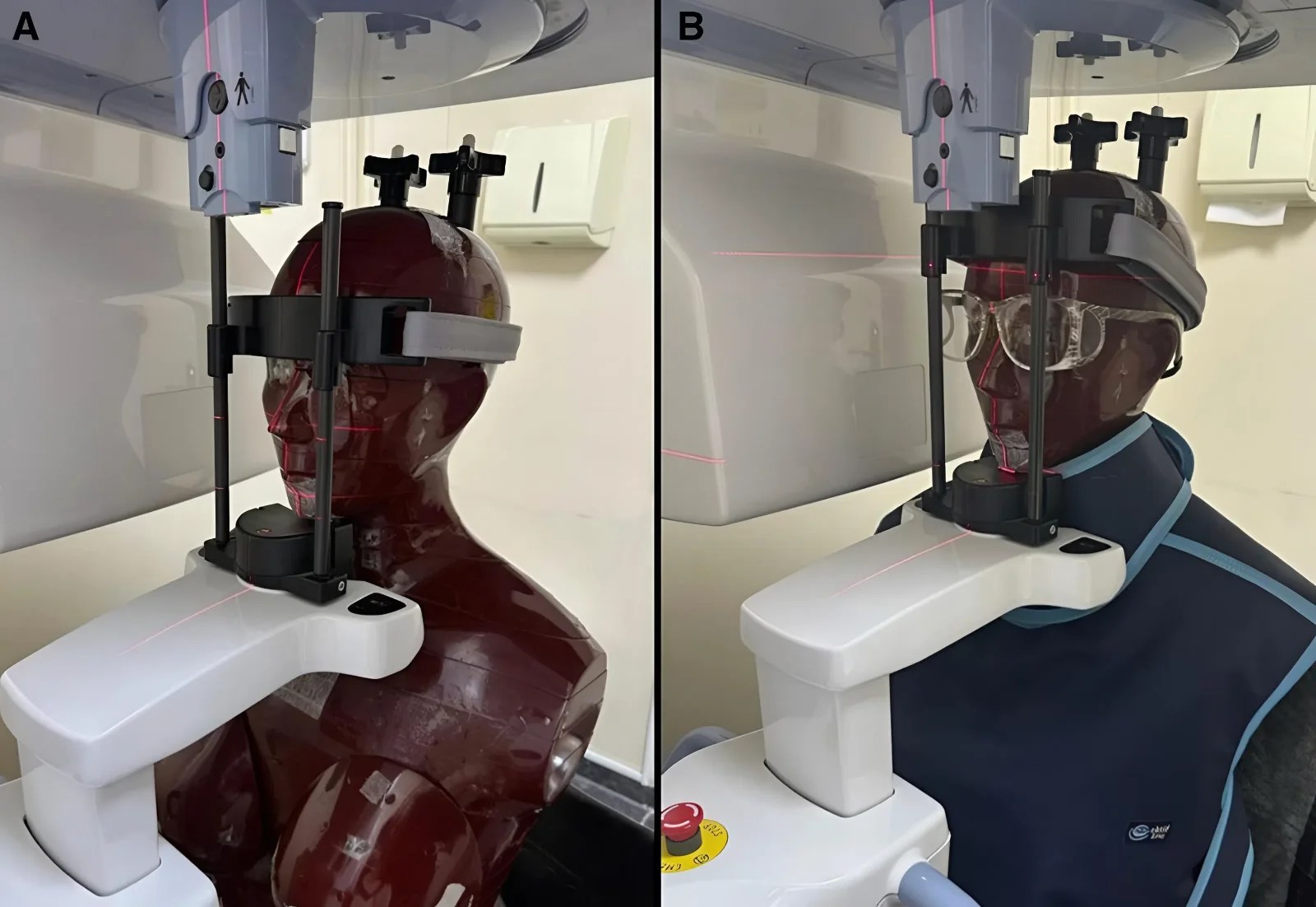
Intraoral Radiography (Periapical and Bitewing Imaging)
Intraoral radiography remains the lowest-dose imaging modality used in routine dental practice. Effective dose for a single digital bitewing typically ranges from 0.3–1.4 µSv, while a four-image bitewing series using rectangular collimation generally falls within 3.4–5.0 µSv.
At these low exposure levels, the additional benefit of thyroid shielding is generally limited, but it may be measurable in specific projections.
In maxillary anterior periapical imaging, vertical beam angulation directs the projected beam toward the cervical region, placing the thyroid closer to the beam path than in many other intraoral projections. Studies have demonstrated measurable thyroid dose reduction in this projection when a collar remains properly positioned outside the imaged region. Hoogeveen et al. (2016)
Despite this potential benefit, practicality determines effectiveness. Thyroid collars may be considered in intraoral imaging only when they:
do not displace the receptor
do not interfere with beam-guiding devices
remain completely outside the field of view
do not increase the likelihood of retakes
If shielding interferes with positioning or image stability, the risk of repeat exposure outweighs any protective benefit.
In practical terms, intraoral radiography is the modality in which thyroid collars are most feasible — but only when placement remains non-disruptive.
Extraoral Imaging (Panoramic and Cephalometric)
Panoramic and cephalometric imaging introduce different positioning constraints. The projected beam passes through the cervical region from multiple angles, and the imaged region includes structures near the neck. As a result, shielding placed around the neck is more likely to enter the field of view.
Typical effective dose ranges include:
Panoramic imaging: approximately 14–30 µSv (digital) and 19–75 µSv (PSP systems)
Cephalometric imaging: approximately 2–10 µSv
Although these exposures remain low, the likelihood of shielding interference is significantly higher than in intraoral imaging.
Several studies have demonstrated modest reductions in measured dose with shielding in panoramic imaging, often in the range of 2–11%. However, this reduction must be weighed against the risk of image obstruction. When shielding obscures the inferior border of the mandible or adjacent structures, the resulting image may be incomplete or non-diagnostic.
In panoramic and cephalometric imaging, shielding is more likely to interfere with image acquisition than to provide meaningful additional protection. For this reason, routine thyroid collar use is generally discouraged in these modalities.
CBCT Imaging
CBCT imaging differs from conventional radiography because radiation exposure varies widely depending on scan parameters rather than shielding alone.
These ranges reflect the dominant influence of field-of-view selection and exposure settings.
Research demonstrates that shielding can reduce absorbed dose in CBCT under controlled conditions. In phantom studies, shielding has produced dose reductions ranging from approximately 26.81% to 52.95%. However, changes in the field of view have produced dose differences ranging from 8.30% to more than 600%, confirming that field-of-view selection remains the primary determinant of radiation exposure.
Shielding also introduces an important technical consideration. When shielding enters or approaches the field of view, it may produce artifacts that compromise diagnostic interpretation or require repeat scanning.
For this reason, shielding is not considered a primary dose-control strategy in CBCT imaging. The most effective controls remain:
justification of the examination
selection of the smallest appropriate field of view
optimized exposure parameters
careful positioning
avoidance of repeat imaging
Shielding may be considered only when it can be confirmed to remain completely outside the scanned region and does not interfere with image reconstruction.
Pediatric Imaging and Dental Radiography During Pregnancy
Pediatric Thyroid Collars in Dental Radiography
Pediatric imaging remains the area where shielding decisions require the most careful judgment. Although routine shielding is not recommended in current U.S. guidance, clinical considerations in children differ from those in adults.
Why Pediatric Imaging Requires Careful Judgment
Children demonstrate greater radiosensitivity than adults, and their longer expected lifespan increases the probability that stochastic effects could manifest over time. In addition, the thyroid gland in children is positioned higher in the neck and occupies a relatively larger portion of the cervical region. This increases the likelihood that the gland may lie closer to the projected beam path, particularly during maxillary imaging or larger-field CBCT studies.
At the same time, pediatric positioning is inherently less predictable. Movement, limited cooperation, and smaller anatomy increase the likelihood of positioning errors and repeat exposures. This balance is critical: shielding that interferes with positioning may increase total exposure rather than reduce it.
Some authors have expressed caution regarding universal discontinuation of shielding in children, emphasizing the radiosensitivity of thyroid tissue and the importance of individualized clinical judgment. While modern dose-reduction strategies remain the primary method of limiting exposure, selective use of thyroid collars may be considered in pediatric patients when placement does not interfere with positioning or image quality. This perspective reinforces the need for thoughtful, case-specific decision-making rather than uniform application of a single rule.
Practical Use of Thyroid Collars in Children
A cautious and clinically defensible approach is to consider thyroid collars in children only when they can be used without interfering with positioning or image quality.
In intraoral imaging, thyroid collars may be considered when they:
remain fully outside the field of view
do not displace the receptor
do not interfere with beam-guiding devices
do not increase retake risk
In panoramic and cephalometric imaging, routine collar use is generally discouraged because shielding is more likely to enter the field of view.
In CBCT imaging, thyroid collars are not considered a primary dose-control method. If shielding has any potential to enter the scanned region or affect image reconstruction, it should not be used.
The most effective protective strategies in pediatric imaging remain:
child-specific exposure settings
selection of the smallest appropriate field of view
accurate positioning
prevention of repeat exposures
These measures consistently produce greater dose reduction than shielding alone.
Dental Radiography During Pregnancy
Pregnancy does not change the fundamental principles of dental imaging, but it increases the importance of careful justification and technique optimization.
What Determines Fetal Dose in Dental Imaging
In dentomaxillofacial imaging, the fetus lies far outside the field of view during intraoral, panoramic, cephalometric, and most CBCT examinations. As a result, fetal exposure is driven primarily by internal scatter originating within the patient, rather than direct beam exposure.
Measured fetal doses during dental imaging are extremely low. Studies evaluating head and neck imaging report abdominal exposures on the order of approximately 1 µGy, a level comparable to a small fraction of typical daily background radiation exposure.
Because exposure is already minimal, shielding placed over the abdomen has little measurable effect on fetal dose.
Why Abdominal Shielding Has Limited Impact
When the imaging field is limited to the teeth and jaws, the amount of radiation reaching the abdomen is extremely small. External shielding cannot meaningfully reduce internally scattered radiation, which represents the primary pathway for distant dose during dental imaging.
For this reason, current recommendations emphasize technique rather than routine abdominal shielding.
Clinical Approach to Imaging Pregnant Patients
The safest and most defensible approach during pregnancy focuses on justification and positioning rather than shielding alone.
Key considerations include:
confirming that imaging will influence diagnosis or treatment
reviewing previous radiographs when available
selecting the smallest appropriate field of view
using optimized exposure settings
preventing repeat imaging
Pregnancy does not prohibit dental imaging when clinically indicated. It reinforces the responsibility to perform imaging intentionally and efficiently.
What Actually Reduces Patient Dose More Than Shielding
In modern dental radiography, the most meaningful dose reductions come from decisions made before and during image acquisition — not from shielding alone. Effective radiation protection depends on technique, equipment selection, and prevention of unnecessary exposure.
Justification and Examination Selection
Dose reduction begins with deciding whether imaging is needed.
Radiographic examinations should be obtained because they are expected to influence diagnosis or treatment planning. Reviewing previous radiographs before acquiring new ones helps avoid unnecessary duplication.
CBCT imaging requires particular discipline. It should not be used routinely or as a screening tool when lower-dose imaging can provide the necessary diagnostic information. When CBCT is indicated, the prescription should be specific to the clinical question.
Justification remains the most effective method of preventing unnecessary radiation exposure.
Field-of-View Selection and Beam Limitation
Restricting the irradiated region to the smallest appropriate field of view consistently produces the greatest reductions in dose.
In intraoral radiography, rectangular collimation reduces radiation exposure by more than 40% compared with round collimation.
In CBCT imaging, field-of-view selection remains the dominant determinant of radiation dose. Reducing the field of view limits the volume of irradiated tissue and produces substantially greater dose reduction than shielding alone.
This reinforces a central principle: dose reduction is most effective when achieved at the source, before exposure occurs.
Exposure Optimization and Positioning
Proper exposure selection and accurate positioning are essential components of dose reduction.
Digital receptors require less radiation than traditional film systems. Exposure parameters should be adjusted according to patient size and examination type, particularly in pediatric patients.
Positioning accuracy also reduces the likelihood of retakes. Stabilization devices, beam-guiding receptor holders, and consistent technique protocols improve first-pass success and reduce cumulative exposure.
A retake represents preventable radiation exposure.
Quality Assurance and Equipment Performance
Equipment reliability plays a direct role in radiation safety. Regular quality assurance ensures that imaging systems perform consistently and produce diagnostic images without unnecessary exposure.
A structured quality program typically includes:
routine equipment inspection
exposure technique charts
verification of system performance
periodic equipment evaluation
For CBCT systems, periodic professional review supports both safety and diagnostic reliability.
Quality assurance is a clinical safety tool — not simply an administrative requirement.
Implementing a Clinic Policy on Patient Shielding
A shielding policy should reflect current professional guidance, applicable regulations, and the realities of clinical workflow.
Routine shielding policies developed decades ago may no longer reflect modern imaging practice. However, regulatory requirements may still mandate shielding in certain jurisdictions. Clinics must confirm applicable state and local requirements before modifying existing protocols.
Guidance vs Regulation: Maintaining Compliance
Professional recommendations guide clinical decision-making, but regulatory requirements govern practice.
Clinic policies should reflect both.
A practical shielding policy typically includes:
discontinuation of routine shielding where regulations allow
availability of shielding when required
selective use when clinically appropriate
documentation of dose-reduction practices
This approach supports regulatory compliance while aligning with current scientific guidance.
Documentation That Supports a Defensible Policy
Documentation demonstrates that radiation safety practices are systematic and consistently applied.
Clinics should maintain:
a written radiation safety program
staff training and licensure records
exposure technique charts
equipment maintenance records
quality assurance documentation
manufacturer instructions
records of periodic equipment evaluation
For CBCT systems, periodic review by a qualified expert supports both regulatory compliance and clinical safety.
Transition Models for Clinics
Clinics updating shielding policies generally adopt one of three approaches:
Discontinue routine shielding where permitted This aligns most closely with current professional guidance.
Maintain shielding available upon request This allows a gradual transition while addressing patient expectations.
Use shielding selectively under defined conditions This approach requires clear criteria to prevent return to routine use.
Regardless of the chosen approach, staff education and consistent implementation remain essential.
Conclusion
Patient shielding in dental radiography is no longer a default precaution. It is a selective decision guided by imaging modality, positioning reliability, and clinical indication.
The most effective protection today comes from justified imaging, controlled field limitation, optimized exposure parameters, and prevention of repeat exposures. These measures consistently reduce radiation exposure more effectively than routine shielding.
Shielding itself is not obsolete. In selected situations — particularly when sensitive tissues lie near the projected beam path and placement does not interfere with positioning — it may provide incremental benefit. However, once shielding compromises positioning or image quality, it ceases to function as protection.
Radiation stewardship in dentistry depends on intentional imaging decisions rather than visible accessories. The responsibility of the clinician is not only to limit exposure but also to ensure that each study acquired is necessary, properly performed, and fully interpreted.
Lead Apron vs Thyroid Collar: What’s the Clinically Meaningful Difference?
They serve different purposes and are not interchangeable.
A thyroid collar may be relevant when the thyroid lies near the projected beam path, such as in selected intraoral projections. An abdominal apron protects tissues that are typically far outside the dental imaging field.
In routine dental imaging, the thyroid collar has greater clinical relevance, while abdominal shielding generally provides minimal additional benefit.
If State Regulations Still Require Aprons, What Should Clinics Do?
Follow the regulation.
Professional guidance informs clinical decision-making, but regulatory requirements govern practice. Clinics should discontinue routine shielding only where regulations allow and maintain shielding availability when required by law or clinical necessity.
A defensible policy reflects both scientific guidance and regulatory compliance.
Does Patient Shielding Reduce Occupational Exposure?
No. Patient shielding does not significantly affect operator exposure.
Occupational protection depends on distance, protective barriers, and positioning outside the projected beam path. Standard recommendations include standing at least 2 meters away or behind a barrier during exposure.
When Is a Thyroid Collar More Likely to Cause a Retake?
When it interferes with positioning or enters the field of view.
This risk is highest in panoramic imaging, cephalometric imaging, and some CBCT studies. In intraoral imaging, shielding should be used only when it remains completely outside the field of view and does not displace the receptor.
Once shielding increases the retake risk, it no longer functions as protection.
How Should Imaging Be Documented During Pregnancy?
Documentation should clearly support justification.
This includes:
the clinical indication
confirmation that prior imaging was reviewed
selection of the smallest appropriate field of view
use of optimized exposure parameters
confirmation that imaging will influence care
Pregnancy does not prohibit imaging when indicated. It requires careful documentation and technique optimization.
Is Thyroid Shielding Ever Justified in CBCT?
Not as a routine measure.
Selective use may be considered only when shielding can be confirmed to remain completely outside the scanned region and when it does not interfere with image reconstruction.
In CBCT imaging, field-of-view selection and exposure optimization remain the primary dose-control strategies.
Does a CBCT Scan Require Full Interpretation Even If Ordered for One Question?
Yes.
When a CBCT volume is acquired, diagnostic responsibility extends to the entire field of view. CBCT interpretation is not limited to the original reason the scan was ordered.
Patient protection includes not only minimizing exposure but also ensuring that all acquired data are fully evaluated.
A practical clinical guide to pediatric dental radiography: when dental X-rays are indicated in children, how to choose the right image, and how to balance diagnostic value with radiation safety.
Invasive cervical resorption is often asymptomatic and easily missed on routine imaging. This article explains how to recognize its key radiographic features, distinguish it from internal resorption, and understand why CBCT is often critical for accurate diagnosis and treatment planning.
Radiation in dentistry isn’t a fear issue — it’s a stewardship issue: every exposure should be justified, dose-optimized, and fully interpreted. This article puts typical dental doses (including CBCT variability) into context and gives dentists a practical framework to image intentionally and document responsibly.